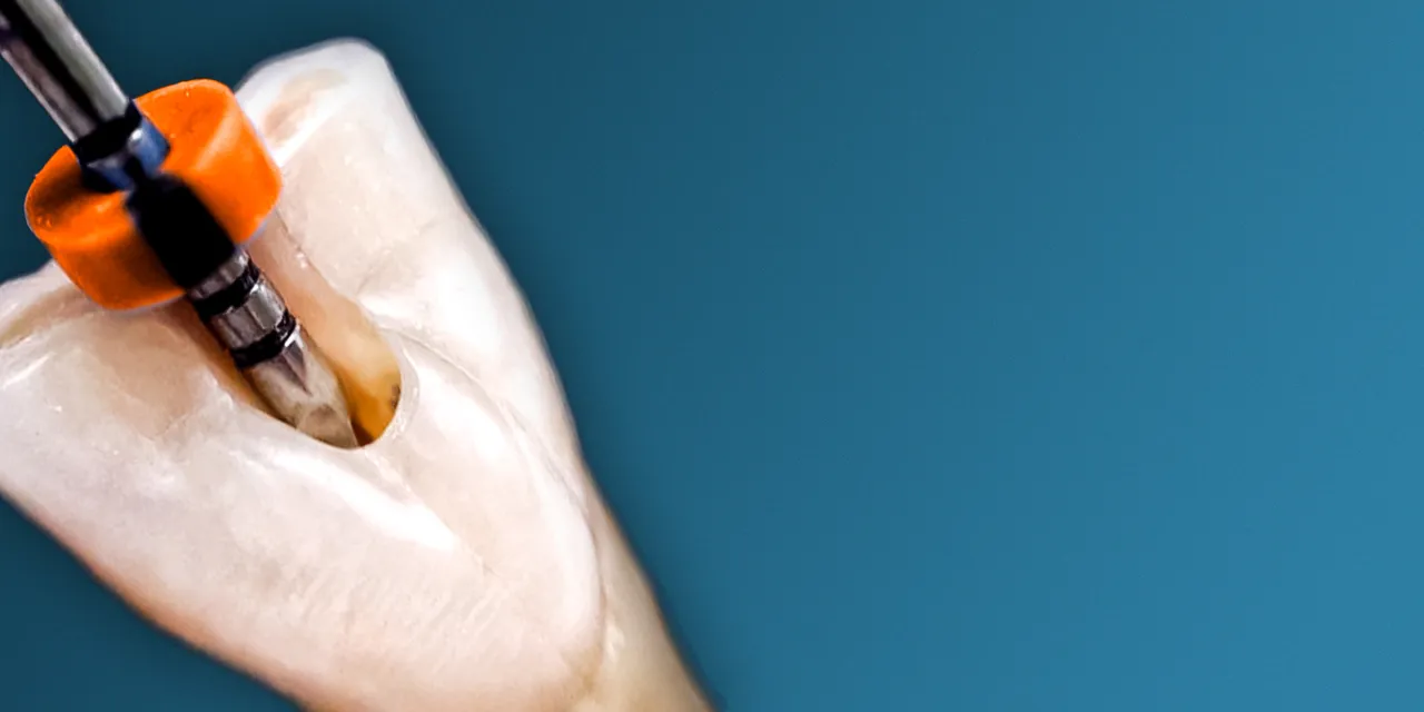
OHI-S Root perforations – diagnosis and treatment
Include: 1 videos + 1 audios + 1 file sub vtt, size: 1 GB
Target Audience: endodontists, restorative dentists, prosthodontists, and general practitioners
Information:
1h 43min
The OHI‑S Root Perforations – Diagnosis and Treatment program is best for endodontists, restorative dentists, prosthodontists, and general practitioners who want structured, training in identifying and managing root perforations. It emphasizes evidence‑based diagnostic protocols, treatment options, and interdisciplinary collaboration to preserve teeth and ensure long‑term success.
👩⚕️ Who Should Enroll
- Endodontists managing complex root canal cases where perforations occur during treatment or due to pathology.
- Restorative dentists & general practitioners performing root canal therapy and restorative procedures who need to recognize and treat perforations.
- Prosthodontists rehabilitating compromised teeth where perforation management is critical for long‑term prognosis.
- Oral surgeons collaborating on surgical repair of perforations in severe cases.
- Residents & fellows in endodontics, restorative dentistry, or prosthodontics seeking advanced training in perforation management.
- Dental technicians supporting restorative workflows where perforation repair impacts prosthetic design.
📚 What You’ll Learn
- Diagnosis protocols: clinical and radiographic methods to identify root perforations.
- Treatment strategies: non‑surgical repair, surgical repair, and use of biocompatible materials (e.g., MTA, bioceramics).
- Prognostic factors: location, size, and timing of perforation repair.
- Interdisciplinary integration: combining endodontic, restorative, and surgical approaches.
- Complication management: preventing infection, periodontal breakdown, and tooth loss.
- Case‑based lessons: real examples of perforation diagnosis and treatment outcomes.
Topics:
– Difficulties in the diagnosis of perforations
– Perforation treatment tools
– Perforation treatment techniques.
Root perforations may occur due to pathological or iatrogenic reasons. Diagnosis is sometimes difficult, but clinical history, radiographic and clinical features usually lead us to understand what is happening. To get a clearer picture, CBCT is an outstanding tool. This information is fundamental to determine the size and location, and hence choose the best treatment option. Several factors contribute to the prognosis, which is in many cases favorable after the correct treatment.


