
National Diagnostic Imaging Symposium™ 2022
$30.00
Details : 118 videos + 2 pdfs, size: 29.3 GB
This Product is shared via google drive download link, So please share your correct Gmail id while placing the order .Please note that there are no CME points or certificate associated with this course Samples for Courses Can be found here : Free Samples Here!
National Diagnostic Imaging Symposium™ 2022
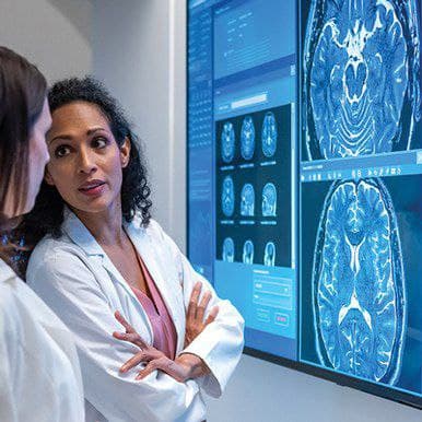
National Diagnostic Imaging Symposium™ 2022 In this comprehensive online CME program, high caliber faculty deliver 119 lectures spanning multiple radiology subspecialties, including Chest, Musculoskeletal, Breast, Gastrointestinal, Ultrasound, Emergency, Genitourinary, Neuroradiology, and Cardiovascular.
World Class CME’s National Diagnostic Imaging Symposium is designed to meet the continuing medical education needs of radiologists — whether in general or specialized care — and radiology residents/fellows who wish to learn to use imaging in a clinical practice setting for screening, diagnosis and staging of injury and disease, including:
- Using CT and MRI to assess injury and disease in the extremities, spine, and cranium
- Applying ultrasound to musculoskeletal, gynecological, obstetrical, cardiovascular, and other organ systems
- Effectively using MDCT and MR imaging to evaluate the abdomen, liver, pancreas, spleen, and bowel
- Improving assessment of genitourinary malignancies, masses, and anomalies using MRI, CT, and US
- Correctly interpreting and staging CT findings in the context of a lung cancer-screening program
And more…
Unique Learning Objectives
At the conclusion of this CME activity, you will be better able to:
- Increase diagnostic capabilities in multiple radiology sub-specialties
- Utilize CT and MRI to assess injury and disease in the extremities, spine, and cranium
- Apply ultrasound to musculoskeletal, gynecological, obstetrical, cardiovascular, and other organ systems
- Effectively use MDCT and MR imaging to evaluate the abdomen, liver, pancreas, spleen, and bowel
- Plan the appropriate imaging of emergency department patients
- Improve assessment of genitourinary malignancies, masses and anomalies using MRI, CT, and US
- Improve diagnostic accuracy in breast imaging and distinguish advantages of the newest breast screening and diagnostic technologies
- Correctly interpret and stage CT findings in the context of a lung cancer-screening program
- Understand how to utilize non-invasive options such as CT, MR, and ultrasound for the evaluation of vascular problems
- Improve skills in MR and US-guided radiology procedures
- Intended Audience
National Diagnostic Imaging Symposium was designed to meet the educational needs of radiologists whether in general or specialized care and radiology residents/fellows who desire to learn, in depth, how to use imaging in a clinical practice setting for screening, diagnosis and staging of injury and disease.
TOPICS / SPEAKERS
Chest Radiology
- Lung Cancer Screening Practice Guidelines – Jared D. Christensen, MD, MBA
- Smoking-Related Lung Disease – Thomas E. Hartman, MD, FACR
- Diffuse Nodular Disease – Jonathan H. Chung, MD
- Imaging of Pleural Disease – Carol C. Wu, MD
- Approach to Fibrotic Lung Disease – Jonathan H. Chung, MD
- Imaging of Cystic Lung Disease – Thomas E. Hartman, MD, FACR
- Approach to Ground Glass Opacities – Carol C. Wu, MD
- Cardiothoracic Manifestations of COVID-19 – Jared D. Christensen, MD, MBA
- CT Pulmonary Angiography – Pearls and Pitfalls – Carol C. Wu, MD
- Imaging of Cardiothoracic Surgical Complications – Jared D. Christensen, MD, MBA
- Non-Malignant Airways Disease – Thomas E. Hartman, MD, FACR
- Signs in Cardiopulmonary Imaging – Jonathan H. Chung, MD
Musculoskeletal Radiology
- Osseous Stress Injuries – Who, What, Where, and Why? – Mark W. Anderson, MD
- Imaging Assessment of Hip Morphology – FAI and Dysplasia – MK Jesse, MD
- Elbow MRI: Case-based Review – Robert D. Boutin, MD
- Post Operative Imaging of the Hip – MK Jesse, MD
- Soft-tissue Masses: Practical Pearls and Pitfalls – Robert D. Boutin, MD
- Tip of the Iceberg Fractures – Little Fractures That Mean Big Trouble – Mark W. Anderson, MD
- Knee MRI: Case-based Review of Misses that Matter – Robert D. Boutin, MD
- Ankle Tendon Pathology – MK Jesse, MD
- MR of the Shoulder – What If It’s Not a Rotator Cuff or Labral Tear? – Mark W. Anderson, MD
Breast MP
- Breast MRI in the Newly Diagnosed Cancer Patient – Debra Monticciolo, MD, FACR
- Optimizing Breast MR Image Quality – Tips for Troubleshooting – Bonnie N. Joe, MD, PhD
- How We Can Improve Upon Routine Breast MRI – Maxine S. Jochelson, MD
- Results of the EA-1141 Abbreviated Breast MRI Trial – Christopher E. Comstock, MD, FACR
- MRI of the Post-Surgical Breast – Reduction, Reconstruction, and Implants – Debra Monticciolo, MD, FACR
- Breast MR Biopsy – From Basics to Tips and Tricks – Bonnie N. Joe, MD, PhD
- Vascular Breast Imaging – MRI vs CEDM – Maxine S. Jochelson, MD
- Workstation Readout and Reporting of Breast MRI – Christopher E. Comstock, MD, FACR
- Breast MRI Teaching Cases – Debra Monticciolo, MD, FACR
- Breast MRI BI-RADS, Challenging and Confusing Scenarios – Bonnie N. Joe, MD, PhD
- Breast MRI in the Neoadjuvant Setting – Maxine S. Jochelson, MD
- Clearly Benign Lesions on Breast MRI – Christopher E. RIComstock, MD, FACR
Gastrointestinal Radiology
- Diffusion Weighted Imaging – Frank H. Miller, MD
- Quantitative CT for Diffuse Liver Disease – Perry J. Pickhardt, MD
- MDCT of Pancreatic Cancer – Elliot K. Fishman, MD
- How to Manage Incidental Hepatic Lesions – Richard M. Gore, MD
- MDCT for Peptic Ulcer Disease – Perry J. Pickhardt, MD
- CT of the Small Bowel: Inflammatory Disease – Elliot K. Fishman, MD
- Challenging Cases in Abdominal Imaging – Perry J. Pickhardt, MD
- CT Evaluation of Gastric Tumors – Elliot K. Fishman, MD
- MR of Liver Metastases – Frank H. Miller, MD
- MDCT of Bowel Obstruction – Richard M. Gore, MD
- MR of Benign Pancreatic Masses – Frank H. Miller, MD
- Complications of Pancreatic Surgery – Richard M. Gore, MD
Ultraso
- US Imaging of IBD – Contributions of Elastography and CEUS – Stephanie R. Wilson, MD, FRCPC
- OB Legal Cases – Lessons Learned – Dolores H. Pretorius, MD, FACR
- Ultrasound of the Painful Scrotum – Thomas C. Winter III, MD
- Advanced Gallbladder Sonography – William D. Middleton, MD
- Problem Solving in the Abdomen with CEUS – Stephanie R. Wilson, MD, FRCPC
- First Trimester Fetal Ultrasound: Basics & Anomalies – Dolores H. Pretorius, MD, FACR
- Ultrasound-Guided Biopsies in the Abdomen – Thomas C. Winter III, MD
- Classic Signs in Abdominal Sonography – William D. Middleton, MD
- Liver Imaging: Why CEUS? – Stephanie R. Wilson, MD, FRCPC
- Fetal Spine: Pearls and Pitfall – Dolores H. Pretorius, MD, FACR
- Molar Pregnancy, RPOC, and Enhanced Myometrial Vascularity – What to Do? – Thomas C. Winter III, MD
- Doppler Evaluation of the Liver – Williaundm D. Middleton, MD
Breast Imaging
- Everything You Wanted to Know About Breast Pain…But Were Afraid to Ask – Daniel Herron, MD
- Lifestyle Changes to Help Prevent Breast Cancer – Daniel Herron, MD
- Optimizing Practice Efficiency with the MQSA Audit – Karla A. Sepulveda, MD
- False-negative Mammography Outcomes – Causes – Edward A. Sickles, MD
- Subtle Mammographic Signs of Malignancy – Edward A. Sickles, MD
- Evaluation and Management of Asymmetries – A Multimodality Approach – Catherine S. Giess, MD
- Advances in Preoperative Localization Techniques – Karla A. Sepulveda, MD
- Whole Breast US Scanning – Techniques and Methods – Daniel Herron, MD
- Challenges and Pitfalls in Tomosynthesis-Guided Biopsies – Catherine S. Giess, MD
- Emerging Technologies: Radiogenomics and Artificial Intelligence – Karla A. Sepulveda, MD
- Risk Assessment Models and Potential Screening Applications – Daniel Herron, MD
- Things That Look Malignant But are Benign and Things That Look Benign But are Malignant – Catherine S. Giess, MD
Emergency Radiology
- Easily Missed Cervical Spine Injuries – Mark P. Bernstein, MD, FASER
- Imaging of Bowel Obstruction: What Really Matters? – Jorge A. Soto, MD
- CT and MR of Intracranial Infections – Wayne S. Kubal, MD
- Imaging Pelvic Ring Trauma – Mark P. Bernstein, MD, FASER
- Imaging Chest Trauma: Avoiding Pitfalls – Jorge A. Soto, MD
- Pearls and Pitfalls of Imaging Orbital Trauma – Krystal L. Archer-Arroyo, MD
- Torso Trauma Bleeding: Pearls and Pitfalls – Mark P. Bernstein, MD, FASER
- Acute Pancreatitis and Acute Cholecystitis – Jorge A. Soto, MD
- CT and MR of Traumatic Brain Injuries – Wayne S. Kubal, MD
Genitourinary Radiology
- Cystic Renal Masses – Applying Bosniak Classification v. 2019 – Stuart G. Silverman, MD, FACR
- MR Imaging for Leiomyomas and Adenomyosis – Iva Petkovska, MD
- Differentiating Benign from Malignant Pelvic Masses – How and When Not to Sit on the Fence – Hebert Alberto Vargas, MD
- Primer on PIRADS for Prostate Cancer – Pointers for Optimal MR Imaging Technique and Interpretation – Mukesh Harisinghani, MD
- CT Urography – How, When, and Why in 2021 – Stuart G. Silverman, MD, FACR
- Mullerian Duct Anomalies on MR Imaging – Iva Petkovska, MD
- MR Imaging of the Treated Female Pelvis: Pearls and Pitfalls – Hebert Alberto Vargas, MD
- Inflammatory Conditions Affecting the GU Tract: Entities You Need to be Aware of – Mukesh Harisinghani, MD
- Pearls and Pitfalls in Gynecological Oncological Imaging – Iva Petkovska, MD
- Imaging Advanced Prostate Cancer: MRI and New (and Not So New) PET Tracers – Hebert Alberto Vargas, MD
- MR Imaging of Pelvic Floor Disorders – Mukesh Harisinghani, MD
- GU Potpourri of Challenging Cases – Stuart G. Silverman, MD, FACR
Advanced Topics in Breast
- Approach to a Mammogram – Beyond the Basics – Michael J. Ulissey, MD, FACR
- Applications of AI in Breast MRI – Karla A. Sepulveda, MD
- Diffusion Weighted Imaging – Is It Ready for Primetime? – Bonnie N. Joe, MD, PhD
- Teaching Points in Patient Management – Michael J. Ulissey, MD, FACR
- Interesting Multi-Modality Breast Imaging Cases – Karla A. Sepulveda, MD
- MRI Safety – Bonnie N. Joe, MD, PhD
- Breast Imagers as Patient Advocates – Karla A. Sepulveda, MD
- Interactive MRI Case Review – Bonnie N. Joe, MD, PhD
Neuroradiology
- Inflammatory and Infectious Disorders of the Spine – Erik Gaensler, MD
- Facial Swelling – Tabassum A. Kennedy, MD
- Cystic Lesions of the CNS – James G. Smirniotopoulos, MD
- Dental Disease – Harprit Bedi, MD
- Lumbar Spine Update – Erik Gaensler, MD
- A Patterned Approach to White Matter Disease – Tabassum A. Kennedy, MD
- Meningioma and Dural Based Masses – James G. Smirniotopoulos, MD
- Temporal Bone Imaging – Harprit Bedi, MD
- Iatrogenic Disorders of the CNS – Erik Gaensler, MD
- Is it a Pituitary Adenoma? – Tabassum A. Kennedy, MD
- Phakomatoses – James G. Smirniotopoulos, MD
- Neck Imaging – Focus on the Thyroid Gland and Lymph Nodes – Harprit Bedi, MD
Cardiovascular Radiology
- CT of Cardiac Masses: Pearls and Pitfalls – Elliot K. Fishman, MD
- Cardiac CT: Imaging of Plaque, Stenosis and Flow – Nikhil Goyal, MD
- Upper Extremity CT Angiography – Geoffrey D. Rubin, MD
- Vascular Applications of Ultrasound Contrast – John S. Pellerito, MD, FACR
- Cardiac MRI: Patterns of Enhancement – Nikhil Goyal, MD
- CT Evaluation of Vasculitis: Key Findings – Elliot K. Fishman, MD
- CT Angiography of the Abdominal Aorta and Its Branches – Geoffrey D. Rubin, MD
- Difficult Carotid Case Review – John S. Pellerito, MD, FACR
Date of Original Release: March 15, 2022
Date Credits Expire: March 15, 2025
Related products
Anesthesiology & pain medicine
$55.00

$40.00
Anesthesiology & pain medicine
$70.00
Anesthesiology & pain medicine
Gulfcoast Ultrasound-Guided Regional Anesthesia : Upper Extremities
$15.00

Courses
$40.00

$40.00
Anesthesiology & pain medicine
Thoracic Radiology Imaging for Clinicians (Chestnet) 2021 (Videos + Quiz)
$50.00
Anesthesiology & pain medicine
$55.00