MRIOnline Mastery Series: Neurofibromatosis Type 1 (NF1) 2021 VIDEOS)
$25.00
This Product is shared via google drive download link, So please share your correct Gmail id while placing the order .Please note that there are no CME points or certificate associated with this course Samples for Courses Can be found here : Free Samples Here!
MRIOnline Mastery Series: Neurofibromatosis Type 1 (NF1) 2021 VIDEOS)
MRIOnline Mastery Series: Neurofibromatosis Type 1 (NF1) 2021: After completing this course, you will be better able to:
- Apply appropriate search patterns to ensure high quality case assessment
- Identify key anatomical landmarks, variations, and abnormalities on imaging
- Accurately interpret advanced imaging cases
- Formulate definitive diagnoses and limited differentials
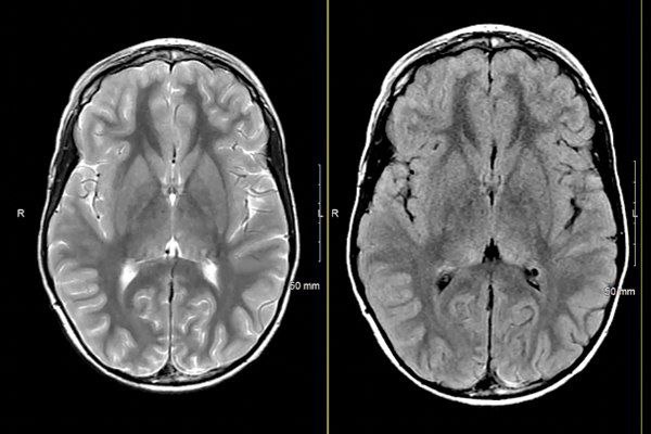
MRIOnline Mastery Series: Neurofibromatosis Type 1 (NF1) 2021 (VIDEOS) In this introductory series on neurocutaneous syndromes, Dr. Asim Choudhri focuses on neurofibromatosis type 1 and its numerous manifestations in the pediatric patient population. This course outlines what findings are clinically significant, how to characterize this disease in reports and the best possible steps for disease management and observation.
Program :
- Faculty and Planning Disclosure
- Introduction to Neurocutaneous Syndromes Part 1 (NF1)
- Chiari Malformation Type 1 in a Patient with NF1
- NF1, With Waxing and Waning Cystic Lesion
- NF1 with Developing Myelin Vacuolization, and Optic Glioma
- NF1 with Suspicious Lesions and Tortuous Optic Nerve
- NF1 with Bilateral Thalamic Lesions and Differential Diagnosis
- Normal Brain MRI in Patients with NF1
- Mild NF1 Phenotype with Sphenoid Wing Dysplasia
- NF1 with High Grade Glioma
- NF1 with Bilateral Optic Nerve Glioma
- Focal Optic Pathway Glioma in NF1
- NF1 with Fusiform Optic Pathway Glioma
- NF1 with Optic Chiasmatic Glioma
- NF1 with Evolution of Optic Nerve Glioma
- NF1 with Optic Pathway Glioma and Moyamoya Disease
- Developing Right Fusiform Optic Glioma
- Glaucoma, an Orbital Manifestation of NF1
- NF1 with a Brain Stem Lesion and Optic Nerve Glioma
- NF1 with Buphthalmos and Orbital Plexiform Neurofibroma
- MRI Appearance of Sphenoid Wing Dysplasia in NF1
- NF1 with Moyamoya Vasculopathy
- Neurocutaneous Syndromes Part 1 (NF1) Summary
Related products
Critical Care - Emergency medicine
2020 Radiology After Five: How to Make Night and Weekend Call a Success!
$15.00

Plastic Surgery
$55.00
Anesthesiology & pain medicine
Gulfcoast Ultrasound-Guided Regional Anesthesia : Upper Extremities
$15.00

Anesthesiology & pain medicine
$35.00

Radiology cousres
$15.00
Critical Care - Emergency medicine
$45.00

Anesthesiology & pain medicine
$45.00