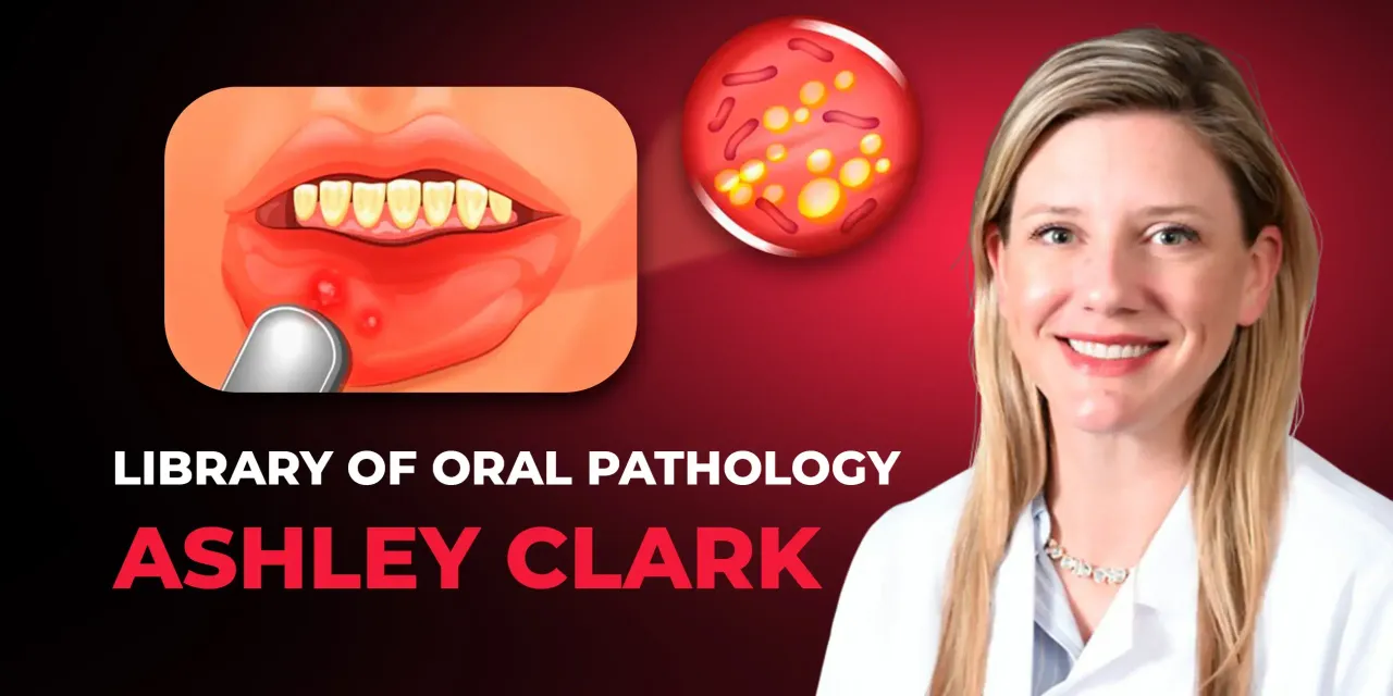
OHI-S Library of Oral Pathology Management of Precancerous and Cancerous Oral Lesions
Include: 4 videos + 4 audios + 4 file sub vtt, size: 1.76 GB
Target Audience: pediatric dentists, periodontists, oral and maxillofacial surgeons, general dentists
Information:
3 lessons + 1 free (3h 25min)
Everything from diagnosis to treatment of the most common precancerous and cancerous oral diseases by Dr. Ashley Clark — A diplomate of the American Board of Oral and Maxillofacial Pathology!
During the course you will learn the protocols of:
– Diagnosis of leukoplakia
– Detection of squamous cell carcinoma of the oral cavity
– Differential diagnosis of ulcerative lesions
– Diagnosis and treatment of salivary glands pathology
– Diagnosis and study of histopathology
– X-ray diagnosis of lesions.
The course covers all questions related to precancerous & cancerous lesions of the oral cavity and protocols for their early diagnosis and treatment.
The OHI‑S Library of Oral Pathology: Management of Precancerous and Cancerous Oral Lesions course is best for oral pathologists, oral medicine specialists, oral surgeons, and general dentists who want structured, training in diagnosing and managing oral precancerous and cancerous conditions. It emphasizes evidence‑based protocols, interdisciplinary collaboration, and practical case management to improve patient outcomes.
👩⚕️ Who Should Enroll
- Oral pathologists & oral medicine specialists focusing on diagnosis and management of oral precancerous and malignant lesions.
- Oral & maxillofacial surgeons performing biopsies, resections, and surgical management of oral cancers.
- General dentists & prosthodontists who need to recognize early signs of precancerous lesions and refer appropriately.
- Periodontists managing gingival lesions with potential malignant transformation.
- Residents & fellows in oral pathology, oral medicine, or oral surgery seeking advanced training.
- Dental hygienists & clinical staff supporting early detection and patient education.
📚 What You’ll Learn
- Precancerous lesions: leukoplakia, erythroplakia, oral lichen planus, and their malignant potential.
- Cancerous lesions: squamous cell carcinoma and other oral malignancies.
- Diagnostic protocols: clinical, histopathological, and adjunctive diagnostic tools.
- Management strategies: surgical, medical, and adjunctive therapies.
- Interdisciplinary collaboration: integrating pathology, surgery, oncology, and restorative dentistry.
- Case‑based lessons: real examples of diagnosis, treatment, and long‑term follow‑up.
Topics:
Lesson 1.Leukoplakia & oral cancer
– Introduction to the classification and the diagnosis protocols of oral and oropharyngeal squamous cell carcinoma
– The etiology of human papillomavirus
– Clinical features of potentially malignant disorders of the oral cavity
– Clinical features of oral dysplasia
– Lesions of leukoplakia
– Thin leukoplakia: hyperkeratosis, acanthosis, carcinoma-in-situ
– Thick leukoplakia: mild dysplasia, mild dysplasia with hyperkeratosis
– Veruciform leukoplakia: severe dysplasia
– Erythroleukoplakia: severe dysplasia, squamous cell carcinoma
– Common locations in which leukoplakia is encountered
– The appropriate treatment plan for patients with leukoplakia
– Lesions of oral squamous cell carcinoma
– Methods on how to help prevent squamous cell carcinoma in your patient population.
Recommended for: Pediatric dentists, Periodontists, Oral and maxillofacial surgeons, General dentists.
Lesson 2.Ulcers of the oral cavity
– Ulcerative conditions of the oral cavity
– The differential diagnosis for ulcerative lesions
– Diagnosis, treatment protocols, and case presentation of desquamative gingivitis
– Differential diagnosis of desquamative gingivitis
– Diagnosis, treatment protocols, and case presentation of lichen planus:
– Reticular form
– Papules form
– Plaque-like form
– Atrophic form
– Ulcerative form
– Bullous form.
– Diagnosis, treatment protocols, and case presentation of mucous membrane pemphigoid (MMP)
– Diagnosis, treatment protocols, and case presentation of Pemphigus vulgaris
– The differences between acute and chronic ulcerative conditions
– Diagnosis, treatment protocols, and case presentation of recurrent aphthous ulceration (RAU): minor and major form
– Diagnosis, treatment protocols, and case presentation of herpes simplex virus and recurrent herpes simplex
– Treatment plan for patients with acute ulcers
– Treatment plan for patients with chronic ulcers
– The follow-up measures for patients with oral ulcerations.
Recommended for: Pediatric dentists, Periodontists, Oral and maxillofacial surgeons, General dentists.
Lesson 3.Common epithelial, mesenchymal, and salivary lesions of the oral cavity
– Pigmented lesions of the oral cavity and their differential diagnosis:
– Amalgam tattoos: diagnosis, treatment protocols, and clinical presentation
– Oral Melanotic macule: diagnosis, treatment protocols, and clinical presentation
– Acquired melanocytic nevus: diagnosis, treatment protocols, and clinical presentation
– Melanoma: diagnosis, treatment protocols, and clinical presentation.
– Human papillomavirus: the difference between low-risk and high-risk HPV strains
– The papillary lesions of the oral cavity and their differential diagnosis:
– Squamous papilloma: diagnosis, treatment protocols, and clinical presentation
– Verruca vulgaris: diagnosis, treatment protocols, and clinical presentation
– Condyloma acuminatum: diagnosis, treatment protocols, and clinical presentation.
– Differential diagnosis of mesenchymal lesions: fibromas and bumps on the gum
– Salivary pathology: mucoceles and xerostomia
– Treatment plan for patients with common oral lesions
– Clinical cases and conclusion.
Recommended for: Pediatric dentists, Periodontists, Oral and maxillofacial surgeons, General dentists.
Lesson 4.Common radiographic oral pathology
– Radiographic features, diagnosis, and management of radiolucent defects:
– stafne defect.
– Radiographic features, diagnosis, and management of multilocular radiolucent lesions:
– ameloblastoma
– central giant cell granuloma
– hemangioma
– odontogenic keratocyst.
– Radiographic features, diagnosis, and management of common radiopaque lesions:
– condensing osteitis
– idiopathic osteosclerosis
– cemento-osseous dysplasia
– focal cemento-osseous dysplasia
– periapical cemento-osseous dysplasia
– florid cemento-osseous dysplasia
– cemento blastoma
– osteoma.
– The difference between lesions with a similar radiographic presentation
– When radiographic lesions do need a biopsy?
– Follow-up measures regarding radiographic pathology
– Syndromes associated with radiographic pathology.
Recommended for: Pediatric dentists, Periodontists, Oral and maxillofacial surgeons, General dentists.


